
WHO defined leukoplakia as “a white patch or plaque that cannot be characterized clinically or pathologically as any other disease”.
Oral leukoplakia is defined as a predominantly white lesion of the oral mucosa that cannot be characterized as any other definable lesion.
It is a Keratotic plaque occurring on mucous membranes and is considered as a premalignant lesion of the oral cavity. Leukoplakia Prevalence is around 2.6% and over 50yrs of age and commonly seen in men.
The etiologic factors of leukoplakia include the following: use of tobacco products (both chewing and smoking forms), alcohol consumption, frequent exposure to extremely hot or cold, spicy, or acidic foods and beverages, use of alcoholic mouth rinses, occlusal trauma, irritation from sharp edges of teeth or prosthetic appliances, exposure to actinic radiation (UV light), infections such as syphilis, presence of Candida albicans, and certain viral infections.
Lip vermilion, buccal mucosa, mandibular gingiva, tongue, floor of the mouth, hard palate, maxillary gingiva, Lip mucosa and soft palate.
Borders may be distinct or indistinct, smoothly contoured or ragged.
Solitary or multiple plaques scattered through the mouth
Floor of the mouth and the lateral borders of the tongue are high-risk sites for
Malignant transformation
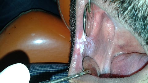
As per the image shown , on inspection a well-defined white patch is observed on both the left and right sides of the buccal mucosa, extending from the lip commissure to the retromolar pad area. On palpation, the lesion is non-scrapable and soft in consistency. Most importantly, the patient has a history of consuming tobacco in the form of bidi, smoking 4-5 bidis per day for the past 5 years.
Homogenous Leukoplakia
A white, well-demarcated plaque with a uniform reaction pattern throughout the entire lesion is observed. There is no red component, and the surface ranges from smooth and thin to a leathery appearance with fissures resembling cracked mud. The lesion is asymptomatic.
Non-Homogenous type
Speckled Leukoplakia: Composed of white and red flecks of fine or coarse variety.
Erythroleukoplakia: Combination of red and white patches, segregation of the red and white components.
Verrucous leukoplakia: Verrucous, papillary (nodular), or exophytic components are present, indicating a more aggressive proliferative pattern. The recurrence rate is high, and such lesions are designated as proliferative verrucous leukoplakia (PVL). PVL is more common in women, with a predilection for the lower gingiva. The malignant potential in PVL is notably high.
High-Risk Leukoplakias
Lesions exhibiting a red component or raised features are typically associated with a higher risk of malignancy. Their presence in high-risk anatomical areas further elevates the concern. A strong correlation exists between tobacco and alcohol use and the development of such lesions, although some cases arise in non-smokers or have an unknown etiology. These lesions are often of a non-reversible type, requiring close monitoring and management. Additionally, microscopic examination may reveal atypia, indicating dysplastic changes and a greater potential for malignant transformation.
I want to emphasize that whenever a patient notices any patches in the oral cavity; they must visit a doctor because early detection will lead to a good prognosis. This is a pre-malignant lesion, and we still have a chance to prevent it from converting into malignancy. I also want to highlight that the majority of the people who are affected are from lower or poor socioeconomic classes and majority are male’s, such as labourers and rickshaw drivers, as they are not very well educated about lesions. Therefore, education and awareness are key steps in preventing pre-malignant lesions which is very important as the cases of these lesions are increasing.
Dr. Nancy Verma BDS, MDS, Senior Lecturer, Santosh Dental College, Ghaziabad, Uttar Pradesh, India
RISK FACTORS & PREVENTION
Preventing leukoplakia involves addressing its risk factors and promoting overall oral health. First and foremost, individuals should avoid tobacco products, including smoking and smokeless forms like chewing tobacco and bidis, as these are significant contributors to leukoplakia development. Limiting alcohol consumption is also crucial, particularly avoiding the use of alcoholic mouth rinses, which can irritate oral tissues. Maintaining good oral hygiene through regular brushing and flossing, along with routine dental check-ups and cleanings, is essential for preventing oral lesions.
Additionally, managing dietary habits by avoiding excessively hot, cold, spicy, or acidic foods and beverages can help reduce irritation of the oral mucosa. It is important to address dental issues, such as sharp edges of teeth or poorly fitting dentures, which may cause chronic irritation. For those at risk due to sun exposure, protecting the lips with balms or by covering them when outdoors is advisable.
Regular screening and prompt treatment of oral infections, such as Candida or viral infections are also important. Raising awareness about the risks of leukoplakia, especially in high-risk populations, can encourage preventive behaviors. Finally, scheduling routine visits to the dentist allows for early detection and management of any abnormal changes in the oral cavity, significantly reducing the risk of developing leukoplakia and promoting better oral health overall.
Narayan TV, Shilpashree S. Meta-analysis on clinicopathologic risk factors of leukoplakias undergoing malignant transformation. J Oral Maxillofac Pathol. 2016 Sep-Dec;20(3):354-361.
Chuang SL, Wang CP, Chen MK, Su WW, Su CW, Chen SL, Chiu SY, Fann JC, Yen AM. Malignant transformation to oral cancer by subtype of oral potentially malignant disorder: A prospective cohort study of Taiwanese nationwide oral cancer screening program. Oral Oncol. 2018 Dec;87:58-63.
Brouns E, Baart J, Karagozoglu Kh, Aartman I, Bloemena E, van der Waal I. Malignant transformation of oral leukoplakia in a well-defined cohort of 144 patients. Oral Dis. 2014 Apr;20(3):e19-24.
Speight PM, Khurram SA, Kujan O. Oral potentially malignant disorders: risk of progression to malignancy. Oral Surg Oral Med Oral Pathol Oral Radiol. 2018 Jun;125(6):612-627.
van der Waal I. Oral leukoplakia: A diagnostic challenge for clinicians and pathologists. Oral Dis. 2019 Jan;25(1):348-349.
Zhu YX. [On the diagnostic problems in oral leukoplakia]. Zhonghua Kou Qiang Ke Za Zhi. 1984 Dec;19(4):203-6.