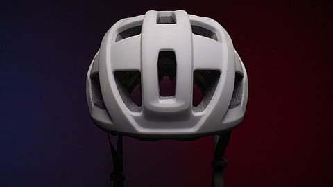

A man walking around a room wearing a helmet that records his brain function would have seemed like science fiction five years ago. Now researchers have designed a lightweight helmet with tiny LEGO-size sensors that scan the brain while a person moves.
The helmet is the first of its kind to accurately record magnetic fields generated by brain activity while people are in motion, reports a new research paper published in NeuroImage. This advance could make it easier to conduct brain scans in young children and individuals with neurological disorders who can’t always remain still in conventional scanners.
Researchers can use the wearable brain scanner, which can be adapted to different head sizes and shapes, to learn more about brain development and what happens in the brains of children and adults who develop neurological conditions such as autism, epilepsy, stroke, concussion, and Parkinson’s disease.
“Unconstrained movement during a scan opens a wealth of possibilities for clinical investigation and allows a fundamentally new range of neuroscientific experiments,” said Niall Holmes, Ph.D., a Mansfield Research Fellow in the School of Physics and Astronomy at the University of Nottingham, who led the research.
How magnetic fields are recorded
When your brain cells (neurons) interact, they generate a small electric current. This electric current produces a magnetic field that can be detected, recorded, and analyzed by sensitive magnetic sensors using a technique called magnetoencephalography (MEG). These sensors must be highly sensitive to detect the low signals that magnetic fields produce.
MEG technology can record both normal and abnormal brain signals every millisecond. The neuronal sources of these magnetic fields are overlaid onto an anatomical image of the brain, allowing clinicians to visualize where and when specific brain activities originate.
MEG systems have evolved to where you can sit and place your head inside them, but they’re bulky and rigid, like an old-fashioned hair dryer where you must keep your head still for a while. In addition, conventional MEG systems have sensors that require cooling at or below freezing temperatures so they can’t be placed directly on your scalp.
Researchers at the University of Nottingham used a new generation of magnetic field sensors called optically pumped magnetometers (OPMs) that operate at room temperature and can be placed close to the head, enhancing data quality. Also, the sensors that are placed in the helmet are flexible, allowing children and adults to move during scanning.
But there is a drawback—OPMs must operate without background “noise” (magnetic fields) that can interfere with the quality of the recording. The researchers had to design a magnetic shielding system that would cancel out or compensate for these magnetic fields.
Designing a matrix coil system
The Nottingham research team constructed a system of electromagnetic coils to shield against the background noise and positioned them on two panels around the participant. Prior research published in Nature shows that eight large coils cancelled the background magnetic fields, but at a fixed position that only allowed small head movements.
Holmes and his team designed a new matrix coil system that features 48 smaller coils on two panels positioned around the participant. The coils can be individually controlled and continually recalibrate to compensate for the magnetic field changes experienced by the moving sensors, ensuring high-quality MEG data are recorded.
“This enables magnetic field compensation in any position, which makes OPM-MEG scans more comfortable for everyone and allows people to walk around,” said Holmes.
The researchers demonstrated the capabilities of the new matrix coil system with four experiments. They first wanted to show that the stationary helmet (not worn by anyone) placed inside the two coil panels could reduce background magnetic fields, which it did. Then a healthy participant wore the helmet, demonstrating that the OPMs recorded his brain function when he moved his head and that the coils cancelled the magnetic fields.
A third experiment used a wire coil as a proxy for brain cell activity because it produces magnetic fields when electric currents are applied. The wire coil attached to the helmet with OPM sensors showed that the matrix coil compensated for motion-related changes, ensuring accurate measurements. The last experiment showed that the helmet worn by a second healthy participant could produce a high-quality recording of brain activity when walking around.
“By taking advantage of recent OPM-MEG technology and designing a new magnetic shielding system, this helmet represents a novel magnetoencephalography approach that could help reveal more about how the brain works,” said Shumin Wang, Ph.D., a program director in the NIBIB Division of Applied Science & Technology (Bioimaging).
The main study limitation was that the matrix coil system must be operated within a magnetically shielded room for the system to function effectively. The room, which measured about 9ft by 9ft in the study, limited the ability of participants to move beyond this controlled setting.
Research applications
A company co-founded by Holmes and his colleagues is selling the OPM-MEG systems, which include a magnetically shielded room, to research centers in North America and Europe to conduct a variety of neuroscientific experiments.
One of the U.S. centers to use the MEG-OPM system is Virginia Polytechnique Institute and State University, which collaborated with the Nottingham team on another study to determine how well the OPM-MEG helmet worked when two individuals each wore one and then interacted. To conduct this proof-of-concept study, two experiments involving social interaction were conducted.
“To really study how the human brain works, we have to embed people in their favorite natural environment—that’s a social setting,” said Read Montague, Ph.D., the principal investigator of the Virginia Tech team and director of the university’s Center for Human Neuroscience Research. The research was published this year in Sensors.
The social exchanges were two participants stroking each other’s hands and then playing a game of ping pong. Both experiments showed that despite large and unpredictable motions by participants, each person’s brain activity was clearly recorded.
Next steps
The company is collecting data to obtain approval from regulatory bodies, including the U.S. Food and Drug Administration, to deploy the system in clinical populations. That process can take up to five years.
This research was funded in part by NIBIB grant (R01EB028772).
Study References:
Holmes, N, et al. Enabling ambulatory movement in wearable magnetoencephalography with matrix coil active magnetic shielding. NeuroImage. (2023) DOI: 10.1016/j.neuroimage.2023.120157
Holmes N, et al. Naturalistic Hyperscanning with Wearable Magnetoencephalography. Sensors. (2023). DOI: 10.3390/s23125454. (MV/Newswise)
