Light used in Imaging Technology for complex aortic repairs

Fiber-optic system in use at UT Southwestern improves safety by decreasing radiation exposure
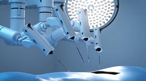
The device, known as Fiber Optic RealShape (FORS) and manufactured by Philips, uses light to visualize blood vessels (Unsplash)
Published on

A new imaging device at UT Southwestern is making complex aortic repairs safer for patients and operating room staff by dramatically reducing their exposure to radiation. The device, known as Fiber Optic RealShape (FORS) and manufactured by Philips, uses light to visualize blood vessels, nearly eliminating the need for X-rays typically used during minimally invasive vascular procedures.

Once this device is placed within a blood vessel, the strain on the optical fibers changes the light’s pathway (Unsplash)
The device, known as Fiber Optic RealShape (FORS) and manufactured by Philips, uses light to visualize blood vessels (Unsplash)
New MRI technique set to transform heart transplant care

As an alternative to conventional imaging, the FORS device uses light traveling through hair-thin optical fibers built in specially designed catheters and wires to display its position and shape inside the body. Once this device is placed within a blood vessel, the strain on the optical fibers changes the light’s pathway. By analyzing how light reflects back along the fibers, a computer algorithm reconstructs and visualizes the full shape of the device.

The result is a real-time, three-dimensional view of the blood vessel that surgeons can overlay on computed tomography images taken before the procedure [Unsplash]
The device, known as Fiber Optic RealShape (FORS) and manufactured by Philips, uses light to visualize blood vessels (Unsplash)
High-Tech Imaging Offers New Way to Detect Signs of Early Glaucoma

The result is a real-time, three-dimensional view of the blood vessel that surgeons can overlay on Computed Tomography images taken before the procedure, providing a roadmap that surgeons can view in any angle to guide the surgery. Fewer X-rays are necessary when using FORS, significantly reducing exposure to patients and staff. (NS/Newswise)

logo
Medbound
www.medboundtimes.com